 --%3E%3Csvg version='1.1' id='Lager_1' xmlns='http://www.w3.org/2000/svg' xmlns:xlink='http://www.w3.org/1999/xlink' x='0px' y='0px' viewBox='0 0 1500 371.7' style='enable-background:new 0 0 1500 371.7%3B' xml:space='preserve'%3E%3Cstyle type='text/css'%3E.st0%7Bfill:%23005EA8%3B%7D%3C/style%3E%3Cg%3E%3Cpath class='st0' d='M237.9 12.6h-89.8v8.9l1.8 0.3c19.6 3 28.5 6.5 32 12.6c1.3 2.2 1.9 4.9 1.9 8.2c0 11.3-7.2 29.4-19 58.8c0 0-28.2 71.2-35.8 90.3c-3.6-9.2-47.8-121.4-47.8-121.4l-1.9-4.7c-3.2-8-6.8-16.7-6.8-24.1c0-3.1 0.6-5.9 2.1-8.4c3.6-6 12.5-9.8 26.3-11.3l1.9-0.2v-9H0v8.9l1.9 0.2c25.4 3 32.5 21.2 41.4 44.2l1 2.6c0 0 70.8 178.5 70.8 178.5h7.2l59.9-150.7l2.8-6.8c17.1-42.2 26.5-65.5 53-67.9l1.9-0.2v-9H237.9z'/%3E%3Cpath class='st0' d='M243.4 87.4l-40.9 14.9v6l1 0.6c18 11 18 14.4 18 27.3v63.2c-0.3 25.2-1 27.6-22.5 31l-1.8 0.3v8.9h76.7v-8.9l-1.8-0.3c-21.6-3.4-22.5-4.5-22.5-31V89.3l-5.5-2.1L243.4 87.4z'/%3E%3Cpath class='st0' d='M216.2 59.3c0 10.4 8.1 18.8 18 18.8c9.9 0 18-8.4 18-18.8c0-10.4-8.1-18.8-18-18.8C224.3 40.5 216.2 49 216.2 59.3'/%3E%3Cpath class='st0' d='M324.9 59.8h-3.7l-45.4 47.4l3.1 4.7h19.8v86.9c0 0 0 5.8 0 6.9c0 13.1 0.5 23.5 7.1 30.1c4.8 4.8 12.3 7.1 23.7 7.1c15.7 0 32.1-3.5 49.9-10.6l1.7-0.7l-2.2-9.2l-2.1 0.6c-5.3 1.5-11.3 2.2-17.8 2.2c-32.1 0-32.1-17.1-32.1-45.4v-68h49.1v-18H327V59.8H324.9z'/%3E%3Cpath class='st0' d='M433.7 87.4l-38.4 14.9v5.8l0.8 0.6c14.4 10.9 14.4 14.4 14.4 27.3v63.3c-0.3 25.2-0.9 27.6-20.9 31l-1.8 0.3v8.8h82v-8.9l-1.9-0.2c-29.2-3.5-29.2-5.6-29.2-31v-33.1l0-1.1c-0.3-17.4 2.2-46.8 22.4-48.8c5.2-0.5 11.7 4.9 16.5 8.8l3 2.4l1.2-0.6c8.4-4.2 17.3-13.1 17.3-22.5c0-10-7.6-17.2-18-17.2c-18.4 0-33.8 17.8-42.1 33.3c0.3-10.5 1-31.3 1-31.3l-5.6-2.1L433.7 87.4z'/%3E%3Cpath class='st0' d='M534.9 163.4c0-31.2 14.1-64.7 45.2-64.7c33.3 0 45.8 42.3 45.8 68.9c0 24.6-11.8 63.7-43.3 63.7C549.7 231.4 534.9 191.6 534.9 163.4 M501.7 165.7c0 45.4 32.3 77.1 78.6 77.1c44.1 0 78.7-34.4 78.7-78.4c0-45.4-32.2-77.1-78.3-77.1C535.7 87.3 501.7 121 501.7 165.7'/%3E%3Cpath class='st0' d='M710.6 0.3l-37.1 15V21l0.9 0.6c14.4 10.9 14.4 14.4 14.4 27.3v150.4c-0.3 25.2-1 27.6-22.5 31l-1.8 0.3v8.9h76.7v-8.9l-1.8-0.3c-21.6-3.4-22.5-4.5-22.5-31V2.1L711.4 0L710.6 0.3z'/%3E%3Cpath class='st0' d='M793.9 87.4L753 102.3v6l1 0.6c18 11 18 14.4 18 27.3v63.2c-0.3 25.2-1 27.6-22.5 31l-1.8 0.3v8.9h76.7v-8.9l-1.8-0.3c-21.6-3.4-22.5-4.5-22.5-31V89.3l-5.5-2.1L793.9 87.4z'/%3E%3Cpath class='st0' d='M766.7 59.3c0 10.4 8.1 18.8 18 18.8c9.9 0 18-8.4 18-18.8c0-10.4-8.1-18.8-18-18.8C774.7 40.5 766.7 49 766.7 59.3'/%3E%3Cpath class='st0' d='M868.3 36.2c-10.5 17.6-12.3 36.1-12.7 53.6c-1.8 1.1-25.3 15.4-25.3 15.4v6.6h25.2v87.6c-0.3 25.2-1 27.6-23.1 31l-1.8 0.3v8.9h82.9v-8.9l-1.9-0.2c-28-3.5-28-5.6-28-31v-87.6h40.4v-18h-40.4V76.3c0-23.1 3.2-61.7 24.9-61.7c14.2 0 27.4 17.4 32.5 24.9l0.6 0.9l1.1 0c6.2 0.2 11.5-1.5 15-4.8c2.9-2.8 4.4-6.5 4.4-11.1c0-17.8-23.4-24.4-38.2-24.4C902.7 0.2 881.9 13.7 868.3 36.2'/%3E%3Cpath class='st0' d='M987.7 98.7c14.1 0 22.2 14 25.3 28c-4.5 0.2-49.3 2.2-54.3 2.4C962.4 114.5 971.9 98.7 987.7 98.7 M925.2 168c0 42.7 29 74.8 67.4 74.8c23.1 0 45.7-15 59.1-39.1l0.9-1.6l-7.9-6.1l-1.3 1.8c-9.7 13.6-20.5 19.7-35 19.7c-14.5 0-27.2-5.5-36.9-16c-10.2-11-15.9-26.2-15.9-42.5c0-1.7 0.1-3.4 0.2-5.1c0 0 0.9-11.4 1.1-14.4h90.5l-0.1-2.2c-1.3-30.5-20.9-50.2-50-50.2C956.2 87.3 925.2 122 925.2 168'/%3E%3Cpath class='st0' d='M1436.4 173.1c0.6-90.8 69.3-98.9 63.2-133.3c-7.7-43.9-149-57.6-253.8-9.8c-36 16.4-67.6 33.7-98.5 57.3c-7.3 5.8-9.5 12.6-10 19.2c0.5 38.6 11.9 74.6 31.1 105.1c6-8.3 12.5-17 18.6-25.1c19.6-25.8 23.2-31.2 57.9-58.8c69.5-55.2 178.1-83.1 186.1-59.1c7.9 23.8-37.2 42.7-37.2 107.2c0 43.2 21.1 73.5 39.6 91.8v0c11 9.9 14.9 6.4 22.3 2.1c0.5-0.3 1-0.5 1.4-0.6c13-9.2 24.9-20 35.4-31.9C1459.4 223.9 1436.2 205 1436.4 173.1'/%3E%3C/g%3E%3Cg%3E%3Cpolygon points='950.2 360.9 937.6 329.4 930.1 329.4 947.5 370.8 952.7 370.8 970.1 329.4 962.7 329.4 '/%3E%3Crect x='980.2' y='329.4' width='6.6' height='41.5'/%3E%3Cpolygon points='994.8 335.2 1006.1 335.2 1006.1 370.8 1012.7 370.8 1012.7 335.2 1024 335.2 1024 329.4 994.8 329.4 '/%3E%3Cpath d='M1056.4 351.5c1.5-1.1 2.6-2.5 3.4-4.2c0.8-1.7 1.2-3.6 1.2-5.7c0-2.5-0.6-4.7-1.7-6.6c-1.1-1.8-2.7-3.2-4.8-4.2c-2-1-4.5-1.5-7.2-1.5h-12.9v41.5h6.6v-16.3h2.9l12.4 16.3h8.3l-13.6-17C1053.1 353.4 1054.9 352.6 1056.4 351.5z M1045.4 349h-4.4v-14.4h4.4c2.8 0 4.9 0.6 6.5 1.9c1.5 1.3 2.3 3 2.3 5.3c0 1.5-0.4 2.8-1.1 3.9c-0.7 1.1-1.7 1.9-3 2.5C1048.8 348.7 1047.2 349 1045.4 349z'/%3E%3Cpath d='M1107 334.9c-1.9-2-4.1-3.5-6.7-4.6c-2.5-1.1-5.2-1.7-8.1-1.7c-2.9 0-5.6 0.6-8.1 1.7c-2.5 1.1-4.7 2.6-6.7 4.6c-1.9 2-3.4 4.3-4.5 6.9c-1.1 2.6-1.6 5.4-1.6 8.4c0 3 0.5 5.8 1.6 8.4c1.1 2.6 2.6 4.9 4.5 6.9c1.9 2 4.1 3.5 6.7 4.6c2.5 1.1 5.2 1.7 8.1 1.7c2.9 0 5.6-0.6 8.1-1.7c2.5-1.1 4.8-2.6 6.7-4.6c1.9-2 3.4-4.3 4.5-6.9c1.1-2.6 1.6-5.4 1.6-8.4c0-3-0.5-5.8-1.6-8.4C1110.4 339.1 1108.9 336.9 1107 334.9z M1105 356.2c-0.7 1.9-1.7 3.5-2.9 4.8c-1.2 1.4-2.7 2.4-4.4 3.2c-1.7 0.8-3.5 1.1-5.5 1.1c-2 0-3.8-0.4-5.5-1.1s-3.1-1.8-4.4-3.2c-1.2-1.4-2.2-3-2.9-4.8c-0.7-1.9-1-3.9-1-6c0-2.2 0.3-4.2 1-6c0.7-1.8 1.7-3.4 2.9-4.8s2.7-2.4 4.4-3.2c1.7-0.8 3.5-1.1 5.5-1.1c2 0 3.8 0.4 5.5 1.1c1.7 0.8 3.2 1.8 4.4 3.2c1.2 1.4 2.2 3 2.9 4.8c0.7 1.8 1 3.9 1 6C1106 352.3 1105.6 354.3 1105 356.2z'/%3E%3Cpolygon points='1131.7 329.4 1125.1 329.4 1125.1 370.8 1147.5 370.8 1147.5 365.1 1131.7 365.1 '/%3E%3Crect x='1158' y='329.4' width='6.6' height='41.5'/%3E%3Cpolygon points='1179.4 370.8 1186 370.8 1186 353 1200.8 353 1200.8 347.2 1186 347.2 1186 335.2 1201.9 335.2 1201.9 329.4 1179.4 329.4 '/%3E%3Cpolygon points='1213.6 370.8 1235.9 370.8 1235.9 365.1 1220.2 365.1 1220.2 353 1235.3 353 1235.3 347.2 1220.2 347.2 1220.2 335.2 1235.9 335.2 1235.9 329.4 1213.6 329.4 '/%3E%3Cpath d='M1282.6 354.2h12.5v0.9c0 1.6-0.3 3-1 4.3c-0.7 1.3-1.6 2.4-2.7 3.3c-1.1 0.9-2.4 1.7-3.9 2.2c-1.4 0.5-2.9 0.8-4.5 0.8c-1.9 0-3.7-0.4-5.3-1.2c-1.6-0.8-3.1-1.9-4.3-3.3c-1.2-1.4-2.2-3.1-2.9-5c-0.7-1.9-1-4-1-6.3c0-2.1 0.3-4.1 0.9-6c0.6-1.9 1.5-3.5 2.7-4.9c1.2-1.4 2.6-2.5 4.3-3.3c1.7-0.8 3.7-1.2 5.9-1.2c2.3 0 4.5 0.4 6.7 1.3c2.2 0.9 4 2 5.5 3.6l3.2-5.1c-1.3-1.2-2.8-2.2-4.5-3.1c-1.7-0.8-3.4-1.5-5.3-1.9c-1.9-0.4-3.8-0.7-5.7-0.7c-3.2 0-6.1 0.6-8.7 1.7c-2.6 1.2-4.8 2.7-6.6 4.7c-1.8 2-3.2 4.3-4.2 6.9c-1 2.6-1.4 5.3-1.4 8.2c0 3.1 0.5 5.9 1.6 8.5c1 2.6 2.5 4.9 4.3 6.9c1.9 2 4 3.5 6.5 4.6c2.5 1.1 5.1 1.6 8 1.6c2.4 0 4.7-0.4 7-1.2c2.3-0.8 4.3-1.9 6.1-3.4c1.8-1.5 3.3-3.3 4.3-5.5c1.1-2.1 1.6-4.5 1.6-7.2V349h-19.2V354.2z'/%3E%3Cpath d='M1335.6 351.5c1.5-1.1 2.6-2.5 3.4-4.2c0.8-1.7 1.2-3.6 1.2-5.7c0-2.5-0.6-4.7-1.7-6.6c-1.1-1.8-2.7-3.2-4.8-4.2c-2-1-4.5-1.5-7.2-1.5h-12.9v41.5h6.6v-16.3h2.9l12.4 16.3h8.3l-13.6-17C1332.3 353.4 1334.1 352.6 1335.6 351.5z M1324.6 349h-4.4v-14.4h4.4c2.8 0 4.9 0.6 6.5 1.9c1.5 1.3 2.3 3 2.3 5.3c0 1.5-0.4 2.8-1.1 3.9c-0.7 1.1-1.7 1.9-3 2.5C1328 348.7 1326.4 349 1324.6 349z'/%3E%3Cpath d='M1386.2 334.9c-1.9-2-4.1-3.5-6.7-4.6c-2.5-1.1-5.2-1.7-8.1-1.7c-2.9 0-5.6 0.6-8.1 1.7c-2.5 1.1-4.7 2.6-6.7 4.6c-1.9 2-3.4 4.3-4.5 6.9c-1.1 2.6-1.6 5.4-1.6 8.4c0 3 0.5 5.8 1.6 8.4c1.1 2.6 2.6 4.9 4.5 6.9c1.9 2 4.1 3.5 6.7 4.6c2.5 1.1 5.2 1.7 8.1 1.7c2.9 0 5.6-0.6 8.1-1.7c2.5-1.1 4.8-2.6 6.7-4.6c1.9-2 3.4-4.3 4.5-6.9c1.1-2.6 1.6-5.4 1.6-8.4c0-3-0.5-5.8-1.6-8.4C1389.6 339.1 1388.1 336.9 1386.2 334.9z M1384.2 356.2c-0.7 1.9-1.7 3.5-2.9 4.8c-1.2 1.4-2.7 2.4-4.4 3.2s-3.5 1.1-5.5 1.1c-2 0-3.8-0.4-5.5-1.1s-3.1-1.8-4.4-3.2c-1.2-1.4-2.2-3-2.9-4.8c-0.7-1.9-1-3.9-1-6c0-2.2 0.3-4.2 1-6c0.7-1.8 1.7-3.4 2.9-4.8c1.2-1.4 2.7-2.4 4.4-3.2c1.7-0.8 3.5-1.1 5.5-1.1c2 0 3.8 0.4 5.5 1.1c1.7 0.8 3.2 1.8 4.4 3.2c1.2 1.4 2.2 3 2.9 4.8c0.7 1.8 1 3.9 1 6C1385.2 352.3 1384.9 354.3 1384.2 356.2z'/%3E%3Cpath d='M1427.6 354.3c0 3.8-0.7 6.6-2.1 8.3c-1.4 1.7-3.6 2.6-6.6 2.6c-3 0-5.2-0.9-6.6-2.6c-1.4-1.7-2.1-4.5-2.1-8.3v-24.9h-6.6v26.7c0 5.1 1.3 9 3.9 11.6c2.6 2.6 6.4 3.9 11.4 3.9c5.1 0 8.9-1.3 11.4-3.9c2.6-2.6 3.8-6.5 3.8-11.6v-26.7h-6.6V354.3z'/%3E%3Cpath d='M1468.5 330.8c-2.1-0.9-4.4-1.4-7.1-1.4h-12.9v41.5h6.6v-16.5h6.3c2.7 0 5.1-0.5 7.1-1.5c2.1-1 3.7-2.5 4.8-4.4c1.2-1.9 1.7-4.2 1.7-6.8c0-2.7-0.6-4.9-1.7-6.7C1472.1 333.1 1470.5 331.7 1468.5 330.8z M1467 345.8c-0.8 1.1-1.8 1.9-3.1 2.4c-1.3 0.5-2.8 0.8-4.5 0.8h-4.4v-14.4h4.4c1.7 0 3.1 0.3 4.5 0.8c1.3 0.5 2.4 1.3 3.1 2.4c0.8 1.1 1.2 2.4 1.2 4C1468.2 343.4 1467.8 344.8 1467 345.8z'/%3E%3Cpolygon points='1492.3 329.4 1490.8 329.4 1489 334.3 1487.1 329.4 1485.7 329.4 1484.9 336.9 1486.3 336.9 1486.8 331.8 1488.6 336.7 1489.4 336.7 1491.2 331.8 1491.7 336.9 1493 336.9 '/%3E%3Cpolygon points='1479.5 330.6 1481.4 330.6 1481.4 336.9 1482.8 336.9 1482.8 330.6 1484.7 330.6 1484.7 329.4 1479.5 329.4 '/%3E%3Cpath d='M664.5 370.9v-41.6h10.8c2.7 0 5.1 0.5 7.1 1.4c2 0.9 3.5 2.3 4.6 4c1.1 1.7 1.6 3.8 1.6 6.3c0 2.4-0.5 4.6-1.6 6.4c-1.1 1.8-2.6 3.3-4.6 4.3c-2 1-4.3 1.5-7.1 1.5h-7v17.6H664.5z M668.3 350.1h6c2.1 0 3.9-0.4 5.4-1.1c1.5-0.7 2.7-1.8 3.6-3.1c0.8-1.3 1.3-2.9 1.3-4.7c0-1.8-0.4-3.4-1.3-4.7c-0.8-1.3-2-2.3-3.6-3c-1.5-0.7-3.3-1-5.4-1h-6V350.1z'/%3E%3Cpath d='M694.4 370.9l16.4-41.6h3.6l16.3 41.6h-4.1l-5.1-13.2h-17.9l-5.1 13.2H694.4z M705 354.3h15.3l-7.6-19.9L705 354.3z'/%3E%3Cpath d='M743 370.9v-41.6h10.8c2.7 0 5.1 0.5 7.1 1.4c2 0.9 3.5 2.3 4.6 4c1.1 1.8 1.6 3.9 1.6 6.3c0 2.2-0.5 4.2-1.4 5.9c-0.9 1.7-2.2 3.1-3.9 4.1c-1.7 1-3.7 1.7-6 1.9l13.8 17.9h-4.9l-13.2-17.5h-4.7v17.5H743z M746.8 350.1h6c2.1 0 3.9-0.4 5.5-1.1c1.5-0.7 2.7-1.8 3.5-3.1c0.8-1.3 1.2-2.9 1.2-4.7c0-2.7-0.9-4.9-2.7-6.4c-1.8-1.5-4.3-2.3-7.5-2.3h-6V350.1z'/%3E%3Cpath d='M787.7 370.9v-38.1H775v-3.5h29.2v3.5h-12.7v38.1H787.7z'/%3E%3Cpath d='M850.7 371.7c-2.9 0-5.6-0.6-8.2-1.7c-2.5-1.1-4.8-2.6-6.7-4.6c-1.9-2-3.4-4.3-4.5-6.9c-1.1-2.6-1.6-5.4-1.6-8.4c0-3 0.5-5.8 1.6-8.4c1.1-2.6 2.6-4.9 4.5-6.9c1.9-2 4.1-3.5 6.7-4.6c2.5-1.1 5.3-1.7 8.2-1.7c2.9 0 5.6 0.6 8.2 1.7c2.5 1.1 4.8 2.6 6.7 4.6c1.9 2 3.4 4.3 4.5 6.9c1.1 2.6 1.6 5.4 1.6 8.4c0 3-0.5 5.8-1.6 8.4c-1.1 2.6-2.6 4.9-4.5 6.9c-1.9 2-4.1 3.5-6.7 4.6C856.3 371.2 853.6 371.7 850.7 371.7z M850.7 368c2.4 0 4.6-0.5 6.6-1.4c2-0.9 3.8-2.2 5.4-3.8c1.5-1.6 2.7-3.5 3.6-5.7c0.9-2.2 1.3-4.5 1.3-7s-0.4-4.8-1.3-7c-0.9-2.2-2.1-4.1-3.6-5.7c-1.5-1.6-3.3-2.9-5.4-3.8c-2-0.9-4.2-1.4-6.6-1.4s-4.6 0.5-6.6 1.4c-2 0.9-3.8 2.2-5.4 3.8c-1.5 1.6-2.7 3.5-3.6 5.7c-0.9 2.2-1.3 4.5-1.3 7s0.4 4.8 1.3 7c0.9 2.2 2.1 4.1 3.6 5.7c1.5 1.6 3.3 2.9 5.4 3.8C846.1 367.6 848.3 368 850.7 368z'/%3E%3Cpolygon points='906.4 332.7 906.4 329.4 885.4 329.4 885.4 370.9 889.2 370.9 889.2 351.8 905.3 351.8 905.3 348.4 889.2 348.4 889.2 332.7 '/%3E%3C/g%3E%3C/svg%3E)
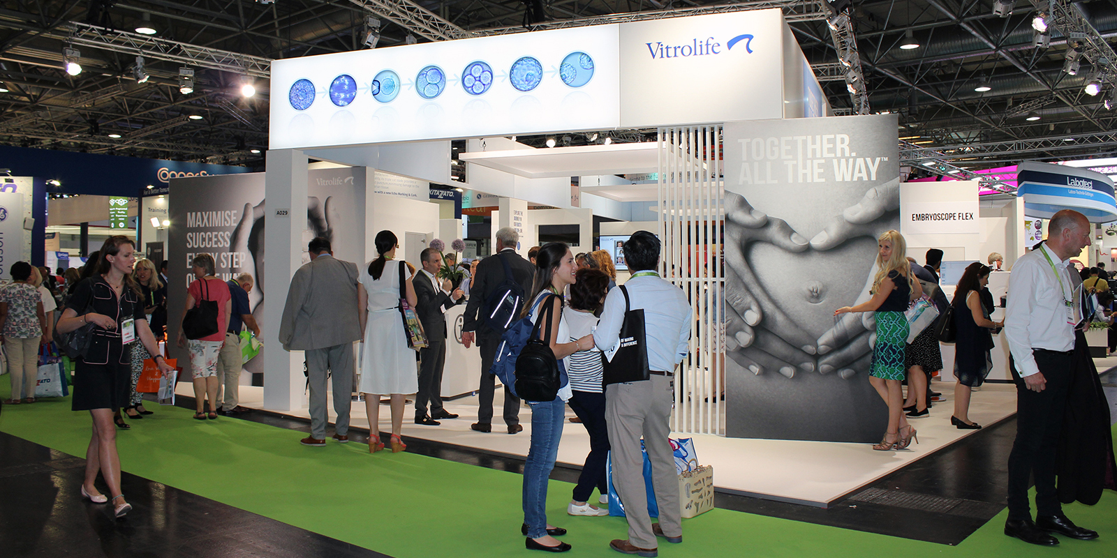
Once again, the biggest event in the field of assisted reproduction, ESHRE, has taken place. No less than 12.003 participants gathered in warm Vienna for updates on the latest science within the field of IVF and of course to meet with old and new friends. As usual the Vitrolife team has put together our thoughts and reflections on some of the scientific content at the meeting. We also provide a new chance to watch our symposium.
Time-lapse technology for embryo selection – now with AI
The clinical and research use of time-lapse technology continues to be of interest at this year’s ESHRE. In all, there were 11 oral and 28 posters reporting use of Vitrolife time-lapse systems. One of the very hot topics is the exploitation of the information provided by time-lapse imaging and development of new methods for embryo selection using artificial intelligence methods for analysing embryo development. Dr. Kragh (O-168) presented how a neural network for grading blastocyst inner cell mass and trophectoderm layers was trained on 7438 embryos by combining information from a sequence of time-lapse images at multiple focal planes. The AI was able to automatically grade ICM and TE with a precision that was at least comparable to experienced embryologists. Automated grading of blastocyst in conjunction with the use of morphokinetics will offer better evaluation consistency and efficiency and improve the accuracy of scoring. Using AI to remove the subjectivity of embryo morphological scoring was also the subject of O-004. Dr. VerMilyea stated that deep learning computer vision AI was trained and validated on static 2D images of Day 5 blastocysts acquired from phase contrast microscopy. It was found that in cases where the embryologist and AI disagreed, the AI was 2.2 times more likely to provide correct fetal heart beat outcome than the embryologist. Across the clinics involved, a 25% relative improvement of accuracy was achieved using AI.
Taking the potential of artificial intelligence even further, the IVY algorithm, which provides a direct estimate of the probability of an embryo leading to viable pregnancy based on full time-lapse sequences, was presented by Dr. Tran at Vitrolife’s symposium. The growing research in the use of artificial intelligence-based methods for eased processes in ART shows that such approaches will improve the speed, precision and workflow of embryo evaluation in the very near future, allowing embryologists to focus on other important tasks in the laboratory. Dr. Ramsing investigated this near future during the A-PART symposium with his talk entitled “The future of embryo evaluation: where is time-lapse and AI taking us?”. During the same symposium another in-depth lecture was given by Dr. Kato from Kato Ladies Clinic, Japan who asked us to “Re-consider the correlation between aneuploidy and embryo morphological and morphokinetic evaluation”.
Ferrick and colleagues (O-169) presented data that human embryos with a higher glucose metabolism have faster blastocoel formation as measured by time-lapse and higher quality grades (morphology score and time-lapse algorithm scores: KIDScore D5 and IVY). Providing a possible physiological reason for their development speed and proposing glucose as a biomarker of embryo viability.
Embryo development patterns for selection & deselection of embryos
Embryo development parameters with potential use for selection/deselection of embryos is always a well-covered topic at ESHRE and this year was no exception. Simonsen et al. (P-209) analysed data for 318 treatments with blastocyst transfer with known implantation outcome. They found that the chance of implantation was significantly higher for embryos that received a high KIDScore Day 5 model score than for embryos with lower scores. Also using KIDScore Day 5, Lim et al. (O-199) were able to show that euploid embryos with higher KIDScore Day 5 values had a greater implantation ratio than low scores.
Direct cleavage (one cell within an embryo dividing into more than two daughter cells) is a known phenomenon and has been well documented to be associated with lower development capacity. It is especially detrimental when the direct cleavage occurs early in the cleavage stage embryo. Shibasaki et al. (P-129) showed that significantly more embryos exhibiting direct cleavage were developed from testicular retrieved sperm compared to ejaculated sperm in their dataset. Reverse cleavage of embryos (cells fusing to set back the number of cells in a cleavage stage embryo) is another well described phenomenon associated with decreased embryo viability. Capdevila et al. (O-117) reported that the ability to reach the blastocyst stage is significantly reduced in reverse cleaving embryos, while the clinical outcome parameters were not significantly affected in embryos that reverse cleaved but achieved blastocyst. This supports previous reports that embryos with poor early development may be rescued by self-repair mechanisms. Rienzi et al. (O-006) showed a classification scheme based on trophectoderm grade (good/poor) combined with time to morula to which may be used to prioritize order of transfer of euploid embryos.
Other interesting investigations of novel embryo development features studies were reported as observed through time-lapse imaging. Dr. Ezoe from Kato Ladies Clinic, Japan (O-113) reported on the formation, duration and distribution of the cytoplasmic halo and its association with abnormal cleavage patterns, Dr. Otsuki (O-116) presented data examining patterns of male and female pronuclei size and kinetic regarding live birth outcome and Eastick et al. (O-005), looked at formation of filopodia in the blastocyst correlated with live birth.
The ESHRE organisation is currently finalising a guideline paper on recommendations for good practice of time-lapse technology. The work from the special interest groups presenting this paper was presented in the ESHRE village by Dr. Giovanni Coticchio. This paper is currently under review and will expectedly be published in its final form later this year.
Focus on genomics and non-invasive preimplantation genetic testing for aneuploidy
There was a real buzz at ESHRE this year around genomics and particularly non-invasive preimplantation genetic testing for aneuploidy (NIPGT-A) by targeting DNA either in blastocoel fluid or spent culture medium. Two years ago, in Geneva, there were several talks that highlighted the problems of maternal contamination, the relatively low efficiency and concordance of the results with those from trophectoderm biopsies at the blastocyst stage and prospects for using NIPGT-A routinely seemed remote. Since then, however, several clinics and companies have been optimising their protocols to detect the small amounts of DNA shed into the medium while avoiding contamination and presented their latest data at the meeting.
Randomised control trial (RCT) of preimplantation genetic testing for aneuploidy (PGT-A) using NGS-based copy number analysis
The first RCT to demonstrate an unequivocal benefit of trophectoderm biopsy and PGT-A used array comparative genomic hybridisation (array CGH) for chromosome copy number analysis, in good prognosis patients below the age of 35 years (Yang et al., 2012). This had a huge impact on the field and led to the development of next generation sequencing (NGS) based chromosome copy number testing methods, including VeriSeq PGS. Dr. Yang and colleagues based in several clinics in southern California have now completed a similar RCT, using NGS-based testing and presented the outcome (O-076). Again, there was a significant improvement in live birth rates per single vitrified-warmed blastocyst transfer from about 47 to 70% (p<0.05) in a young group of patients (average age 32 years). Furthermore, since only single embryos were transferred in both arms of the study there were no twin or higher order pregnancies.
Progress with non-invasive testing for aneuploidy (NIPGT-A)
Two alternative approaches to non-invasive PGT-A are currently being explored. The first minimally invasive approach is to sample blastocoel fluid (‘blastocentesis’) in the process of collapsing the blastocoel cavity ahead of vitrification and has been pioneered by Luca Gianaroli’s group in Bologna, Italy. Intriguingly, in their latest paper with senior clinical embryologist, Christina Magli as first author (who was inaugurated as the new President of ESHRE in the opening ceremony) (Magli et al., 2019), they demonstrated much better concordance rates with aneuploid trophectoderm biopsies but failed to amplify any DNA from the euploid blastocysts. This suggests that the DNA in the blastocoel cavity may be derived from aneuploid cells selectively undergoing programmed cell death or apoptosis. If confirmed in larger independent studies, this raises the intriguing possibility that the presence or absence of DNA could provide a simple test for the euploid/aneuploid status of the embryo. In contrast, Sunney Xie’s group (Harvard, Boston and Beijing, China) among others, are pioneering completely non-invasive testing using spent culture medium find DNA in both euploid and aneuploid embryos and even claim that this DNA may be more representative because they believe it is derived from both the inner cell mass from which the fetus is derived and the trophectoderm (Huang et al., 2019).
Interestingly, one group from Create Fertility and the University of Toronto, Toronto, Canada, have combined samples from both sources by collapsing the blastocoel cavity before collecting the medium and then performing trophectoderm biopsy after the blastocyst had re-expanded (Kuznyetsov et al., 2018) and Dr. Madjunkova presented their results (O-201). They measured nanogram quantities of DNA in the samples from both good and poor-quality blastocysts following whole genome amplification and informative NIPGT-A results were obtained from 70/71 (99%) of trophectoderm samples and 63/71 (89%) of combined blastocoel fluid/culture medium samples using NGS-based testing (VeriSeq PGS). Furthermore, there was complete concordance between the NIPGT-A and conventional PGT-A trophectoderm samples. They suggest that collecting combined samples in this way provides consistent results and could be used, for example, as a backup if no results are obtained from the first biopsy eliminating the need to rebiopsy the embryo and with further validation used routinely for aneuploidy testing.
Frozen embryo transfers
Dr. Terho (O-036) reported similar childhood growth patterns between frozen embryo transfer children and fresh embryo transfer children, indicating the safety and feasibility of embryo freezing. Terho et al. studied the growth of children, until five years of age, born after frozen embryo transfer (FET) compared to fresh embryo transfer (Fresh ET). It has previously been shown that birth weight of FET children are higher than fresh ET children and spontaneously conceived children. However, the knowledge on the childhood growth patterns of these children is still very limited. This study indicates that the childhood growth is similar between FET and fresh ET children after adjustments for age at measurement, maternal BMI and parity. There were no significant differences in birth weight or length. If a frozen embryo transfer is performed it might be best to schedule it sooner rather than later. Dr. Li (O-068) presented data demonstrating that immediate frozen embryo transfer following a stimulated IVF cycle had a significantly higher ongoing pregnancy rate than delayed frozen embryo transfer. Dr. Kataoka et al. (O-037) provided data that suggests that the trophectoderm grade influences pregnancy rate and gender following frozen embryo transfer. The Kato group (Ueno et al. (O-305) presented data on live birth from low/minimal stimulation cycles using day 7 blastocysts that were transferred after vitrification/warming in natural ovulatory cycles (synchronised to D5). A multivariable logistic regression analysis showed that female age, blastocyst expansion time and diameter influenced live birth following D7. Live birth rate (LBR) was significantly lower in D7 compared to D5 and D6. Using cut-off values for D7 blastocysts (confirmed time of blastulation >143 hrs, expansion time < 19hrs and diameter >210 µm), the adjusted LBR on D7 was still lower than on D5, but not different from D6. Thus, the cut-off values may be a good selection criterion for D7 blastocysts which may be subjected to PGT, and that the add-on of time-lapse may help to find optimal cut-off parameters.
Improving implantation
An et. al. (O-302) assessed in a multicenter retrospective cohort study if the transfer of single euploid blastocysts at 120 ± 12 hrs post was superior compared to out-of-window (asynchronous) transfers. 1079 D5/6 blastocyst transfers were performed in both natural ovulatory (n=878) or in ovulation induction cycles (n=201). LBR was significantly higher following transfer in the optimal time window. A significant difference was also observed in a sub-evaluation looking at top or good quality embryos, concluding that the optimal time for transfer is independent of embryo quality and that an optimal implantation window for vitrified/warmed blastocyst transfers does exist. According to Dr. Kitaya (O-022) repeated implantation failure could be attributed to the microorganism (microbiota) in the endometrial fluid and vaginal secretions. In women where lactobacillus dominated the microbiota in the endometrial fluid, during the receptive phase, the clinical outcomes were more favourable. Endometrial scratching has been proposed as a way to improve receptivity. Van Hoogenhuijze and colleagues (O-023) presented data on a multicenter, non-blinded randomized control trial with patients that had already undergone a failed IVF cycle. Endometrial scratching did not impact clinical pregnancy or live birth rates. Messaoudi et al (O-025) have developed a RT-qPCR based test (Adehsio RT) to predict receptivity and embryo implantation by identifying genes involved in the implantation window and the maternal-embryo dialogue. Expression profiles were determined following 50 biopsies during a natural cycle in the proposed implantation window. The expression profiles were compared between those that resulted in a successful pregnancy and those that did not. Autologous co-culture of cells derived from biopsies and embryos was also performed. Samples were analysed using microarrays to identify potential biomarkers. Ten genes were selected as biomarkers and clinical validation was performed on 130 biopsies from IVF-patients with a known pregnancy outcome.
Clinical studies - raising the bar
The conclusion of the presenter of O-218, a statistician from the Cochrane group (Wilkinson), is that clinical studies in the IVF field are too heterogenous and there is a need for improved study design, reporting and standardization of the outcome parameter definitions. The current heterogeneity makes it extremely difficult to perform meaningful meta-analysis of a given treatment. For example, less than half the randomised control trials reporting live birth did so correctly by including all randomized patients or only removing those patients violating the protocol. To improve the quality of clinical studies Fertility and Sterility offer a registered report scheme that allows study plans to be peer-reviewed based on their importance and methodology prior to initiation. Dr. Duffy (O-219) talked about efforts to prioritise unanswered questions relating to IVF treatment. The Priority Setting Partnership for Infertility (Oxford University) and the Cochrane Gynaecology and Fertility group. By engaging a number of health care professionals, researchers and patients representing different geographies COMMIT (Core Outcome Measures for Infertility Trials) has developed core outcomes. Both presentations highlighted the importance of study design and homogenous outcome measures/reporting, which will result in increasing knowledge, improved treatments and reduce research waste in terms of both time and financial resources.
Watch our Scientific Symposium "Meeting patient needs: Shortening the time to a healthy live birth"
This year we had the pleasure of listening to Dr. Tran talking about the future of embryo selection with time-lapse AI algorithms. Prof. Alan Handyside took us through preimplantation genetic testing for aneuploidy for embryo selection. And finally, Prof. Gardner gave insights into how you can optimise transfer outcome with the use of EmbryoGlue. All three talks centering around how we can help meet patient needs of shortening the time to a healthy live birth.
If you missed the symposium or simply would like to see it again, just click below.

Topics: IVF community insights